When your pet is sick or injured you want help, and you want answers quickly. Providing the right kind of help and determining an appropriate treatment plan is contingent on a proper diagnosis. At Oradell Animal Hospital we are able to diagnose with precision – offering the most sophisticated and advanced digital diagnostic tools, coupled with our highly specialized team of doctors and support staff. Depending upon the situation, our veterinarians will recommend the appropriate course of diagnostic action for each patient’s symptoms and needs.
If your pet is not a patient of Oradell Animal Hospital but is referred for a diagnostic procedure, our team will collaborate with your primary care veterinarian to ensure a seamless level of care.
Radiography
The most common and recognized imaging technique in human and veterinary medicine. Using electromagnetic waves, xray imaging creates black and white pictures of internal structures within the body. X-rays are used for various purposes and depending on the part of the body, radiation is blocked or absorbed differently. Radiography produces just 1 image at a time. Often times multiple images are taken at different angels for better diagnostic interpretation.
- Denser tissues (like bone) block most of the radiation and appear bright white
- Soft tissue, such as muscle and fat, absorb more radiation and appear darker
Magnetic Resonance Imaging
An MRI machine uses magnetic fields to create detailed, high-quality, cross sectional images of a body part or region. Anesthesia is required as the patient and area of study need to be perfectly still, but an MRI is otherwise non-invasive and does not cause any harm to your pet.
Diseases/Conditions Diagnosed with MRI:
-
Brain Diseases
- Tumors
- Infarcts – lack of blood to an area of the brain
- Abscesses
- Inflammation of the brain covering (meninges)
-
Spinal Disorders
- Herniated disc
- Intervertebral Disc Disease (IVDD)
- Stenosis – narrowing of the vertebral column
- Cervical Disc Disease
- Nerve root impingement
- Spinal tumors
Sonography
Sonograms, also knows as ultrasounds, have become an essential imaging tool in veterinary medicine and can greatly enhance a veterinarians diagnostic and therapeutic capabilities. It is fast, non-invasive, and the results are available immediately. Using ultrasonic sounds waves, images of internal body structures that are not detectable via other diagnostic tools, such as tissue, organs and even blood flow, are generated digitally. Sonography is typically used to identify and monitor chronic abdominal and cardiac conditions, but is also utilized in acute situations such as trauma or locating an obstructing foreign body. Sonograms are painless, non-invasive, and do not require anesthesia.
Indications for Ultrasound:
- Pregnancy
- Urinary Tract Disease
- Gastrointestinal Disease
- Endocrine Disease
- Pancreatitis
- Neoplasia
- Fever Of Unknown Origin (FUO)
- Immune Mediated Diseases
- Extent Of Tumor Invasion
- Presence Of Tumor Metastases
- Ultrasound Guided Biopsies
Indications for Echocardiography:
- Any evidence of congenital or acquired cardiac disease to determine the structure and function of the heart
- Heart murmur
- Arryhthmia, Gallop
- Breed specific predisposition for heart disease
- Pre-anesthetic Evaluation
Computed Tomography
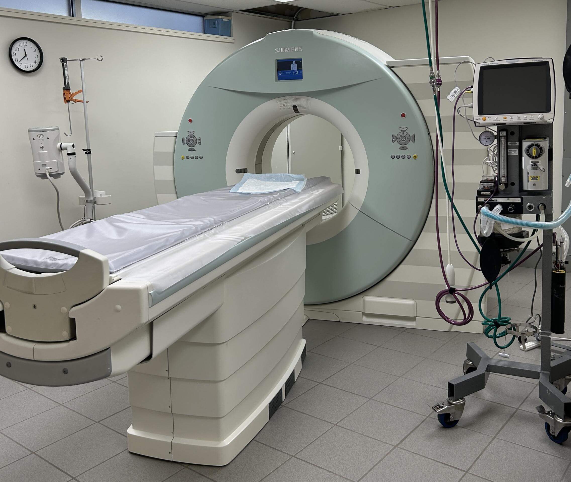
A CAT scan is used to take a closer look at a particular organ, muscle, bone, or other internal structure by combining multiple x-rays into a 3D model. A CT scan renders “slices” of your pet’s body in exceptional detail. Cat scans are especially beneficial when it comes to visualizing the precise location of a tumor or other abnormality and its relationship to neighboring structures.
Indications for CT:
- Inner Ear Infections
- Tumors
- Nasal Disease
- Tooth Decay And Abscesses
- Trauma and Poly-Trauma – when multiple organs and systems are damaged

Endoscopy
An endoscope, a flexible tube with a light and high resolution camera attached to it, is inserted into the body and provides a clear picture of various internal structures. Scoping is minimally invasive and biopsies can be obtained for further evaluation and testing. Anesthesia is required for these procedures.
Endoscopic Techniques:
- Arthroscopy – visualize, diagnose and treat problems inside a joint
- Bronchoscopy – view the internal airways (trachea, esophagus, bronchi and bronchioles in the lungs
- Colonoscopy – detect changes or abnormalities in the large intestine (colon) and rectum
- Cystoscopy – view the urethra and bladder
- Endoscopy – examine the digestive tract (esophagus, stomach and small intestine), used in emergency situations to alleviate life-threatening esophageal foreign bodies
- Rhinoscopy – exam the inside of the nose, nasal passages, sinus cavity and throat
Fluoroscopy
Fluoroscopy allows us to visualize the inside of the body in real time, by taking multiple still x-rays to create an motion x-ray. Fluoroscopy is used for a variety of procedures, but most specifically during tracheal stent placement and to measure the blood flow through the coronary arteries during cardiac catheterization. Many times a contrast agent is administered to provide further detail.
